Untitled Document. Рис. 69. Соединения позвонков

Рис. 69. Соединения позвонков.
Сагиттальный распил на уровне двух поясничных позвонков. 1-тело позвонка; 2-студенистое ядро межпозвоночного диска; 3-передняя продольная связка; 4-фиброзное кольцо межпозвоночного диска; 5-верхний суставной отросток поясничного позвонка; 6- задняя продольная связка; 7-межпозвоночное отверстие; 8-желтая связка; 9-суставная капсула дугоотростчатого (межпозвоночного) сустава; 10-межостистая связка; 11-надости-стая связка.
Fig. 69. Соединения позвонков.
Сагиттальный распил на уровне двух поясничных позвонков. 1-corpus vertebrae; 2-nucleus pulposus; 3-ligamentum longitudinale anlerius; 4-anulus fibrosus; 5-processus articularis superior vertebrae lumbalis; 6-ligamentum longitudinale posterius; 7-foramen intervrete-brale; 8-ligamentuni flavum; 9-capsula articularis art. zygapophysealis (intervertebralis); 10-liganienttim interspinale; 1 l-ligamentum supraspinale.
Fig. 69. Vertebral joints.
Sagittal section at the level of two lumbar verterbrae. 1-vertebral body; 2-nucleus pulposus of intervertebral'disc; 3-anterior longitudinal ligament; 4-anulus fibrosus of intervertebral disc; 5-supe-rior articular process of lumbar vertebra; 6-posterior longitudinal ligament; 7-intervertebral foramen; 8-ligamenta tlava; 9-articular capsule of zygapophysial (intervertebral) joint; 10-interspinous ligament; 11-supraspinous ligament.

Рис. 70. Атланто-затылочный и атланто-осевые суставы (articulatio atlanto-occipitalis et articulationes atlantoaxialis).
Вид спереди (со стороны позвоночного канала). Твердая мозговая оболочка и покровная мембрана удалены. 1-затылочная кость; 2-мродольные пучки крестообразной связки атланта; 3-канал подъязычного нерва (подъязычный канал); 4-крыловидная связка; 5-суставная щель атланто-затылочного сустава; 6-крестообразная связка атланта; 7-суставная шельбоко-вого атланто-осевого сустава; 8-продольные пучки крестообразной связки атланта; 9-капсула атланто-затылочного сустава.
Fig. 70. Articulatio atlan-tooccipitalis et articulatio atlantoaxialis. Вид спереди (со стороны позвоночною канала). Твердая
мозговая оболочка и покровная мембрана удалены. 1-os occipitalc; 2-tasciculi longitixlinales ligament! cruciformis atlantis; 3-canalis hypoglossalis; 4-ligamentum alare; 5-cavitas articularis articulaiionis atlantooc-cipitalis; 6-ligamentum cruciforme atlantis; 7-cavitas articularis aniculationis atlanto-axialis lateralis; 8-fasciculi longiludinales ligamemi cruciformis atlantis; 9-capsula articu-lationis atlantooccipitalis.
Fig. 70. Atlanto-occipital and atlanto-axial joints. Anterior aspect (from vertebral canal).
Dura mater and tectorial membrane are removed. 1-occipital bone; 2-longitudinal ands of cruciate ligament of atlas; 3-hypoglossal canal; 4-alar ligaments; 5-articular cavity of atlanto-occip-ital joint; 6-cruciate ligament of atlas; 7-articular cavity of lateral atlanto-axial joint; 8-longitudinal bands ol'cruciate ligament ot'atlas; 9-capsule of atlanto-occipital joint.

Рис. 71. Срединный атланто-осевой сустав (articulatio atlantoaxialis mediana). Вид сверху. 1-задний бугорок атланта; 2-позвоночное отверстие; 3-верхняя суставная ямка (атланта); 4-отверстие поперечного отростка атланта; 5-поперечная связка атланта; 6-срединный атланто-осевой сустав (задняя часть); 7-зуб осевого позвонка; 8-срединный атланто-осевой сустав (передняя часть); 9-передний бугорок атланта; 10-крыловидная связка.
Fig. 71. Articulatio atlantoaxialis mediana. Вид сверху. 1-tuberculum posterior atlantis; 2-foramen vertebrale; 3-fovea articu-laris superior (atlantis); 4-foramen transversarium atlantis; 5-ligamen-tum transversum atlantis; 6-articulatio atlantoaxialis mediana (pars posterior); 7-dens axis; 8-articulatio atlanto-axialis mediana (pars anterior); 9-tuberculum anterior atlantis; 10-ligamentum alare.
Fig. 71. Median atlanto-axial joint. Superior aspect. I-posterior tubercle of atlas; 2-vertebral foramen; 3-superior articular facet (of atlas); 4-foramen transversarium of atlas; 5-transverse ligament of atlas; 6-median atlanto-axial joint (posterior part); 7-dens of axis; 8-median atlanto-axial joint (anterior part); 9-anterior tubercle of atlas; 10-alar ligament.

Рис, 72. Срединный атланто-осевой сустав (articulatio allantoaxialis mediana). Сагиттальный разрез. 1-передняя продольная связка; 2-тело осевого позвонка; 3-попе-речная связка атланта; 4-атланто-осевой сустав (задняя часть); 5-атланто-осевой сустав (передняя часть); 6-зуб осевого позвонка; 7-передняя дуга атланта; 8-связка верхушки зуба; 9-передняя атланто-затылочная мембрана; 10-покровная мембрана; 11-по-звоночная артерия; 12-твердая мозговая оболочка; 13-затылоч-ная кость(чешуя); 14-задняя луга атланта; 15-1 и II спинномозговые нервы.
Fig. 72. Articulatio atlantoaxialis mediana. Сагиттальный распил. 1-ligamentum longitudinale anterius; 2-corpus axis; 3-ligamentum transversum atlantis; 4-articulatio atlanto-axialis (pars posterior); 5-articulatio atlanto-axialis (pars anterior); 6-dens axis; 7-arcus anterior atlantis; 8-ligamentiim apicis dentis; 9-memhrana atlanto-occipitalis anterior; 10-inembrana tectoria; 11-arteria vertebralis; 12-dura mater; 13-osoccipitale (squama); 14-arcus posterior atlantis; 15-nervi spinales.
Fig. 72. Median atlanto-axial joint (sagittal section). 1-anterior longitudinal ligament; 2-body of the axis; 3-transverse ligament of the atlas; 4-atlanto-axial joint (posterior part); 6-dens of the axis; 7-anterior arch of the atlas; 8-apical ligament of the dens; 9-ante-rior atlanto-occipital membrane; 10-tectorial membrane; 11-vertebral artery; 12-dura mater; 13-occipital bone (squamous part); 14-posterior arch of atlas; 15-1 and II spinal nerves.

Рис. 73. Ребсрно-позвоночные суставы
(articulationescostovertebralis).
Поперечный разрез через позвоночный столб на уровне VI грудного позвонка.
1-тело VI грудного позвонка; 2-лучистая связка головки ребра; 3-сустав головки ребра; 4-головка ребра; 5-медиальная реберно-иомеречная связка; 6-бугорок ребра; 7-реберно-поперечный сустав; 8-попсрсчпый отросток VII грудного позвонка; 9-позвоноч-ное отверстие; 10-желтая связка; II-задняя продольная связка; 12-верхний суставной отросток VI грудного позвонка; 13-ребер-но-поперечный сустав. 14-реберно-поперечная связка: 15-медиальная реберно-поперечная связка; 16-лучистая связка головки ребра; 17-передняя продольная связка.
Fig. 73. Articulationes costovertebrales. Поперечный разрез черепа через позвоночный столб на уровне
шестого [рудного позвонка.
1-corpus vertebrae lumbalis (LVI); 2-ligamentum radiatum capitis costae; 3-articulatio capitis costae; 4-caput costae; 5-liganientum costo-transversarium mediale; 6-tuberculum costae; 7-articulatio costotrans-versaria; 8-processus transversus vertebrae thoracicae (Th VII); 9-fora-men vertebrale; 10-ligamentum flavum; ll-ligamentum longitudinale posterius; 12-processus articularis superior vertebrae thoraeicae (ThVl); 13-articulatio costotransversaria; 14-ligamentum tuberculi costae; 15-ligamenium costotrans-versarium mediale; 16-ligamentum radiatum eapitis costae; 17-ligamentum longitudi-nale anterias.
Fig. 73. Costovertcbral joints.
Transverse sectoin of vertebral column at the level VI thoracic vertebra, l-body of the VI thoracic vertebra; 2-radiate ligament of head ofrib; 3-joint ol'head ofrib; 4-head ofrib; 5-medial costotransverse ligament; 6-tubercle ot rib; 7-coslotransverse joint; 8-transverse process ol the VII thoracic vertebra; 9-vertebral foramen; 10-ligamenta flava; 11-posterior longitudinal ligament; 12-superior articular process of the VI thoracic vertebra; 13-costotransverse joint; 14-costotransversal ligament; I5-medial costotransverse ligament; 16-radiatc ligament of head ofrib; 17-postcrior longitudinal ligament.

Рис. 74. Связки реберно-позвоночных суставов. Вид сбоку. 1-верхняя реберная ямка; 2-верхний суставной отросток грудного позвонка; 3-реберная ямка поперечного отростка; 4- межпоперечная связка; 5-лучистая связка головки ребра; 6-реберно-попе-речные отверстия; 7-верхняя реберно-поперечная связка; 8-межпозвоночные диски; 9-передняя продольная связка.
Fig. 74. Связки реберно-позвоночных суставов. Вид сбоку. 1 -fovea costalis superior; 2-processus articularis superior vertebrae tho-racicae; 3-fovea costalis transversa; 4-ligamentum intertransversarium; 5-ligamentum radiatum capitis costae; 6-loramina costotransversaria; 7-ligamentum costotransversarium superior; 8-disci intervertebrales; 9-ligamentum longitudinale anterius.
Fig. 74. Ligaments of the costovertebral joints. Lateral aspect. 1-superior costal facet; 2-superior articular process of thoracic vertebra; 3-tranverse ligament; 4-intertransverse ligament; 5-radiate ligament of head of rib; 6-costo-transverse foramen; 7-superiorcostotransverse ligament; 8-intervertebral disce; 9-anterior longitudinal ligament.

Рис. 75. Связки позвоночника и реберно-позвоночных суставов. 1-связка бугорка ребра; 2-надостистая связка; 3-желтая связка; 4-реберно-ноперечная связка; 5-латеральная реберно-иопереч-ная связка; 6-межпоперечные связки; 7-внутренняя межреберная мембрана.
Fig. 75. Связки позвоночника и реберно-позвоночных суставов, l-ligamentum tuberculi costae; 2-ligamentum supraspinatum; 3-liga-mentum llavum; 4-ligamentum costotransversarium; 5-ligamenta cos-totransversaria lateralia; 6- membrana intercostalis interna.
Fig. 75. Ligaments of vertebral column and costovertebral joints. 1-ligament of tubercle of rib; 2-supraspinas ligament; 3-ligamenta flava; 4-costotransverse ligament; 5-lateral costotransverse ligament; 6-internal intercostal membrane.
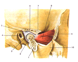
Рис. 76. Височно-нижнечелюстной сустав (articulalio temporomaiKlibularis). Сагиттальный разрез. 1-суставной (мышелковый) отросток нижней челюсти; 2-головка нижней челюсти; 3-суставиая капсула; 4-наружный слуховой проход; 5-суставной (внутрисуставной) диск; 6-нижнечелюстная ямка; 7-суставной бугорок; 8-латеральная крыловидная мышца; 9-височный отросток скуловой кости (отрезан); 10-венечный отросток нижней челюсти.
Fig. 76. Articulatio temporornandibularis. Сагиттальный разрез. 1-processus articularis (condylaris) mandibulae; 2-caput mandibulae; 3-capsula anicularis; 4-porus acusticus extemus; 5-discus articularis; 6-fossa mandibularis; 7-tuberculum articulare; 8-m.pterygoideus later-alis; 9-processus temporalis ossis zygomatici; 10-processus coro-noideus.
Fig. 76. Tenporomandibular joint (sagittal section). 1-articular (condylar) process of mandible; 2-head of mandible; 3-articular capsule; 4-external acoustic opening; 5-articular disc; 6-mandibular fossa; 7-articular tubercle; 8-lateral pterygoid muscle; 9-temporal process of zygomatic bone (it is cut); 10-coronoid process of mandible.

Рис. 77. Связки височно-нижне-челюстного сустава.
Вид с медиальной стороны.
1-латеральная связка (височно-нижнечелюстного сустава); 2-капсула височно-нижнечелюстного сустава; 3-клиновидно-нижнечелюстная связка; 4-шило-нижнечелюстная связка; 5-от-верстие нижней челюсти; 6-скуловая дуга; 7-КЛИНОВЩЩая пазуха; 8-гипофизарная ямка (турецкого седла).
Fig. 77. Связки височно-нижнечелюстного сустава.
Вид с медиальной стороны.
1-ligamentum laterale (articulatio temporomandibularis); 2-capsula articulationis temporomandibularis; 3-ligamentum sphenomandibu-lare; 4-ligamentum stylomandibulare; 5-foramcn mandibulae; 6-arcus zygomaticus; 7- sinus sphenoidalis; 8-fossa hypophysialis.
Fig. 77. Ligamenta of tempotamandibular joint.
Medial aspect.
1-lateral ligament (of temporomandibular joint); 2-capsula of tem-poromandibular joint; 3-sphenomandibular ligament; 4-styIomandibu-lar ligament; 5-mandibular foramen; 6-zygomatic arch; 7-sphenoidal sinus; 8-hypophysial fossa (of sella turcica).
К.фмшшьш iiTiac анатом, чело г.
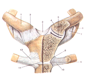
Рис. 78. Грудино-ключичный сустав (articulatiostemoclavicularis). Вид спереди. На левой стороне препарата сустав вскрыт
фронтальным разрезом.
1-ключица (правая); 2-передняя грудино-ключичная связка; 3-межклюмичная связка; 4-грудинный конец ключицы; 5-внут-рисуставной диск (фудино-ключичного сустава); 6-первое (I) ребро; 7-реберно-ключичная связка; 8-фудино-реберный сустав (11-го ребра); 9-внутрисуставная грудино-реберная связка; 10-хрящ 11-го ребра; 11-синхондроз рукоятки фудины; 12-лучис-тая фудино-реберная связка.
Fig. 78. Грудино-ключичный сустав. Вид спереди. На левой стороне препарата сустав вскрыт фронтальным разрезом. 1-clavicula (dextra); 2-ligamentum sternoclaviculare anterius; 3-liga-mentum interclaviculare; 4-extremitas sternalis claviculae; 5-discus articularis (articulatio sternoclavicularis); 6-costa (I); 7-ligamentum costoclaviculare; 8-articulatio sternocostalis (II); 9-ligamentum sternocostalis intraarticulare; 10-cartilago costae (II); ll-synchondrosis manubrii sterni; 12-ligamentum sternocostale radiatum.
Fig. 78. Sternoclavicular joint. Anterior aspect. Frontal section of joint at left. 1-clavicle (right); 2-anterior Sternoclavicular ligament; 3-interclavicular ligament; 4-sternal end ot'clavicle; 5-articular disc (ol'sternoclavicular joint); 6-1 rib; 7-costoclavicular ligament; 8-sternocostal joint (of the II rib); 9-intra-articular sternocostal ligament; 10-cartilage of the II rib; 11-manubriosternal synchondrosis; 12-radiate sternocostal ligament

Рис. 79. Акромиально-ключичный суетав (articulatio acromio-clavicularis) и плечевой суставы
(articulatio humeri). Вид спереди.
1-акромион; 2-акромиалыю-ключичный сустав (акромиально-ключичная связка); 3-клювовидно-акромиальная связка; 4-клю-вовидный отросток; 5-клювовидно-ключичная связка; 6-ключи-ца; 7-верхняя поперечная связка лопатки; 8-лопатка; 9-капсула плечевого сустава; 10-плечевая кость; 11-сухожилие длинной головки двуглавой мышцы плеча; 12-подлопаточная мышца; 13-клювовидно-плечевая связка.
Fig. 79. Акромиально-ключичный и плечевой суставы.
Вид спереди.
1-acromion; 2-articulatio acromioclavicularis (ligamentum acromio-claviculare); 3-ligamentum coracoacromiale; 4-processus coracoideus; 5-ligamentum coracoclaviculare; 6-clavicula; 7-ligamentum transver-sum scapulae (superius); 8-scapula; 9-capsula articulationis humeri; 10-humerus; 11-tendo m. bicipitis brachii (caput longum); 12-m.sub-scapularis; 13-ligamentum coracohumerale.
Fig. 79. Acromioclavicular and shoulder joints. Anterior aspect, l-acromion; 2-acromioclavicular joint (acromioclavicular ligament); 3-coraco-acromial ligament; 4-coracoid process; 5-coracoclavicular ligament; 6-clavicule; 7-superior transverse scapular ligament; 8-scapula; 9-capsula of shoulder joint; 10-humerus; 11-tendon of the long head of biceps brachii; 12-subscapularis; 13-coracohumeraI ligament.
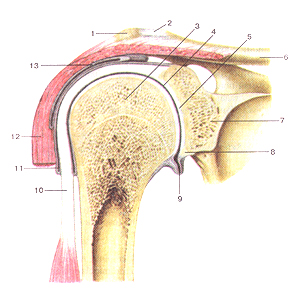
Рис. 80. Плечевой сустав (articulatio huineri).
Фронтальный разрез.
1-акромион; 2-акромиалыю-ключичный сустав; 3-голова плечевой кости; 4-суставная щель плечевого сустава; 5-суставная впадина лопатки; 6-ключица; 7-лопатка; 8-суставная губа; 9-подмы-шечный карман суставной полости; 10-сухожилие длинной головки двуглавой мышцы плеча; 11-синовиальное влагалище сухожилия длинной головки двуглавой мышцы плеча; 12-дельто-видная мышца. 13-поддельтовидная сумка.
Fig. 80. Articulatio humeri. Фронтальный разрез. 1-acromion; 2-articulatioacromioclavicularis; 3-caput humeri; 4-cav-ilasarlicularisarticulatioiiis humeri; 5-cavitasglenoidale (scapulae); 6-clavicula; 7-scapula; 8-labrum glenoidale; 9-recessus subaxilaris cavi-tae articularis; 10-tcndo m.bicipitis brachii (caput longum); 11-vagina synovialis tendinis m.bicipitis brachii (caput longum); 12-m.del-toideus; 13-bursa subdelloidea.
Fig. 80. Shoulder joint. Frontal section.
1-acromion; 2-acromioclavicularjoint; 3-head of humerus; 4-articular cavity of shoulder joint; 5-glenoid cavity (of scapula); 6-clavicule; 7-scapula; 8-glenoid labrum; 9-subaxillar recess of articular cavity; 10-tendon of long head of the biceps brachii; 1 l-inlertubercular tendon sheath (of biceps brachii); 12-deltoid; 13-subdeltoid bursa.

Рис. 81. Локтевой сустав (articulatiocubiti).
Вид спереди.
1-плечевая кость; 2-сустав-ная капсула; 3-медиальный надмыщелок плечевой кости; 4-локтевая коллатерать-ная связка; 5-кольцевая связка лучевой кости; 6-су-хожилие двуглавой мышцы плеча; 7-косая хорда; 8-локтевая кость; 9-лучевая кость; 10-лучевая коллатеральная связка; 11-лате-ральный надмышелок.
Fig. 81. Articulatio cubiti.
Вид спереди.
1-humerus; 2-capsula articu-laris; 3-epicondylus medialis humeri; 4-ligamentum collat-erale ulnaris; 5-ligamentum anulare radii; 6-tendo m.bicipitis brachii; 7-chorda obliqua; 8-ulna; 9-radius; 10-ligamentum collaterale radi-ale; ll-epicondylus lateralis.
Fig. 81. Elbow joint. Anterior aspect.
1-humerus; 2-articular capsula; 3-medial epicondyle of humerus; 4-ulnar collateral ligament; 5-anular ligament of radius; 6-tendon of biceps brachii; 7-oblique cord; 8-ulnar (bone); 9-radius; 10-radial collateral ligament; 11-lateral epicondyle.

Рис. 82. Локтевой сустав и соединения костей предплечья (полость локтевого
сустава вскрыта).
I-плечевая кость; 2-головка мыщелка плечевой кости; 3-блок плечевой кости; 4-по-дость локтевого сустава; 5-бугристость локтевой кости; 6-тело локтевой кости; 7-меж-костная мембрана; 8-дистальныЙ луче-лок-тевой сустав; 9-тело лучевой кости; 10-косая хорда; 11-сухожилие двуглавой мышцы плеча (отрезано); 12-колыдевая связка лучевой кости: 13-головка лучевой кости; 14-капсула локтевого сустава.
Fig. 82. Локтевой сустав и соединения костей предплечья (полость локтевого
сустава вскрыта).
l-humcrus; 2-capitulum humeri; 3-trochantcr liumeri; 4-cavitas articularis; 5-tuberositas ulnae; 6-corpus ulnae; 7-membrana interossea ante-brachii; 8- articulatio radioulnai'isdistalis; 9-cor-pus radii; I0-chorda obliqua; I l-tendo in.bicipi-tis brachii (отрезано); 12-ligamentum anulare radii; 13-caput radii; 14-capsula articularis.
Fig. 82. Elbow joint and joints of forearm (cavity of the elboww joint is discovered). 1-humcrus; 2-capitulum humeri; 3-trochlea of humcrus; 4- articular cavity (of elbow joint); 5-tuberosily of ulna: 6-body of the ulna; 7-intcrosscus membrane of forearm; 9-body of radius: 10-obliquc cord; II-tendon of biceps brachii (is cut); 12-anular ligament of radius; 13-head of radius; 14- articular capsula of elbow joint.

Рис. 83. Луче-запястный сустав (articulartio radiocarpea);
связки и суставы кисти, правой. Ладонная сторона. 1 -локтевая кость; 2-дистальный луче-локтевой сустав; 3-локтевая коллатеральная связка запястья; 4-гороховидная кость; 5-горохо-видно-крючковая связка; 6-гороховидно-пястная связка; 7-крю-чок крючковидной кости; 8-ладонные за мястно-пястные связки; 9-ладонные пястные связки; 10-глубокие поперечные пястные связки; 11-пястно-фаланговый сустав V пальца (вскрыт); 12-межфаланговые суставы V пальца; 13-сухожилие мышцы -глубокого сгибателя пальцев; 14-фиброзное влагалище сгибателей III пальца кисти; 15-коллатеральные связки пястно-фалангового и межфалангового сусгавов 1 пальца кисти; 16-запя-стио-пястный сустав I пальца кисти; 17-головчатая кость; 18-лу-чевая связка запястья; 19-лучевая коллатеральная связка; 20-ладонная луме-запястная связка; 21-полулунная кость; 22-лу-чевая кость; 23-межкостпая перепонка предплечья.
Fig. 83. Aniculatio radiocarpea. Связки и суставы кисти,правой. Ладонная сторона, l-ulna; 2-articulatio radioulnaris distalis; 3-ligamcntum collaierale саф1 ulnare; 4-os pisiforme; 5-ligamentum pisohamatum; 6-ligamen-tum Р18оте1асафсит; 7-hamulus ossis hamati; 8-ligamenta car-pometacarpea palmaria; 9-ligamenta metacarpea palmaria; 10-liga-mcnta тс!асафса transversa prolunda; 11-articulatio caфophalangea (вскрыт); 12-articulationes 1шефЬа1апЕеае manus; 13-tendo m.flex-oris digitorum profundi; 14-vagina tendinis musculi flexorum digito-rum (III); 15-ligamenla collateralia; 16-articulatio сафоптаасафеа; 17-oscapitatum; 18-^атепШтсаф1 radiatum; 19- ligamemum col-laterale carpi radiale; 20-ligamentum сафоте!асафеа palmare; 21-os lunatum; 22-radius; 23-membrana interosseaanterbrachii.
Fig. 83. Radota^al joint, ligaments and joints of wrist, right.
Palmar view.
l-ulna; 2-distal radioulnar joint; 3~ulnar collateral сафа! ligament; 4-pisilbnTi bone; 5-pisohomote ligament; 6-р15оте(асафа1 ligament; 7- hanuilus of hamate bone: 8-palmar
сафате!асафа1 ligament; 9-palmar metacarpal ligament; 10-deep transverse metacarpal ligaments; I 1 -metacarpopha-longeal joint of V finger (dissect); 12-inter-pholongcal joints of V finger; 13-tendon of llexor ciigitorum pro-fundus; 14-tendinous sheath of flecar digito-rum (HI); 15-collaler-al ligaments of metacarpophalangeal and interphalangeal joints of thumb; 16-сафоте1асафа1 joint of thumb; 17-capitate bone; 18-radiale carpal ligament: 19-radial collateral ligament of wrist joint: 20-palmar radiocarpal ligament: 21-lunate bone; 22-radius; 23-interosseous membrane of forearm.

Рис. 84. Луче-запястый сустав (articulanio radiocarpea); суставы и
связки кисти, правой; Разрез во фронтальной плоскости. I-пястные кости; 2-межкостные межпястные связки; 3-запяст-но-пястныс суставы; 4-запястно-пястный сустав 1 пальца кисти; 5-многоугольная кость; 6-трапециевидная кость; 7-головчатая кость; 8-лучевая коллатеральная связка запястья; 9- ладьевидная кость; 10-луче-запястный сустав; 11-полулунная кость; 12-луче-вая кость; 13-локтевая кость; 14-мешкообразное углубление дис-тального луче-локтевого сустава; 15-дистальный луче-локтевой сустав; 16-внутрисуставиой диск луче-запястного сустава; 17-локтевая коллатеральная связка запястья; 18-трехграннан кость; 19-гороховидная кость; 20-межкостные межзапястные свяжи; 21-крючковидная кость; 22-межпястные суставы.
Fig. 84. Articulatio radiocarpea; суставы и связки кистиб правой.
Разрез во фронтальной плоскости.
1-ossa metacarpea; 2-ligamenta interossea intermetacarpea; 3-articu-lationes сафопшасафеае; 4-articulatio carpometacarpea pollicis; 5-os irape/.ium; 6-os trapezoium; 7-os capitatum; 8-ligamentum collat-erale саф1 radiale; 9-os scaphoideum; 10-articulatio radiocaфeunl; 11-os lunatum; 12-radius;13-ulna; 14-recessussacciformis; 15-articu-latio гадюсафеит distalis; 16-discus articularis; 17-Ugamentum col-laterale carpi ulnare; 18-os triquelrum; 19-os pisiformc; 20-ligamenta т!егсафеа interossea; 21-os hamatum; 22-ligamenta ймегсафеа.
Fig. 84. Кад4осафа1 joint, ligaments and joi nls pf right wrist.
Frontal plane.
1-те1асафа1 bones; 2-interosseous те!асафа1 ligaments; 3-car-роте!асафа1 joints; 4-сафоте1асафа1 joint of thumb; 5-tropezium; 6-trapezoid; 7-capitatc; 8-radial сафа! collateral ligament; 9-scaphoid; 10-гаШосафа1 joint; ll-lunate; 12-radius; 13-ulna; 14-sacciform recess of distal radioulnar joint; 15-distal radioulnar joint; 17-и1пагсафа1 collateral ligament; 18-triquetrum; 19-pisiform; 20-interosseous inter-сафа! ligaments; 21-hamate; 22-1п1егсафа1 ligaments.
Дата добавления: 2014-12-11 | Просмотры: 1453 | Нарушение авторских прав
1 | 2 | 3 | 4 | 5 | 6 | 7 | 8 | 9 | 10 | 11 | 12 | 13 | 14 | 15 | 16 | 17 | 18 | 19 | 20 | 21 | 22 | 23 |
|