|
АкушерствоАнатомияАнестезиологияВакцинопрофилактикаВалеологияВетеринарияГигиенаЗаболеванияИммунологияКардиологияНеврологияНефрологияОнкологияОториноларингологияОфтальмологияПаразитологияПедиатрияПервая помощьПсихиатрияПульмонологияРеанимацияРевматологияСтоматологияТерапияТоксикологияТравматологияУрологияФармакологияФармацевтикаФизиотерапияФтизиатрияХирургияЭндокринологияЭпидемиология |
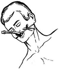
Fig. 8. Laryngoscopy made by means of a laryngoscope with a curved bladeIt is necessary to see that the external end of the blade do not press on the incisors of the upper jaw while the epiglottis is lifted upward, since this may lead to their injury. The tube is passed into the trachea under visual control and moved forward until the whole inflated cuff disappears behind the vocal cords and if it is absent the tube is moved for 3-5 cm lower a true glottis (Fig. 9). In order to control a proper intubation it is necessary: to press on the patient's chest, in so doing, a stream of air coming out of the intubation tube should be defined; to carry out the inflation of gas mixture into the patient's lungs by means of anesthetic apparatus bag, that must lead to the uniform expansion of the chest, appearance of respiratory murmurs over the both lungs and presence of air stream coming out of the intubation tube during the expiratory phase. While passing the tube into one of the primary bronchi on the opposite side on the surface of the chest, respiratory murmurs are not auscultated. While passing the tube into the esophagus in the period of artificial respiration an inflation of epigastric region, absence of respiratory murmurs over the pulmonary surface, development of cyanosis are observed, at the moment of the air inflation into the tube a characteristic murmur ("gurgle") is heard. In separate cases one cannot succeed in carrying out the intubation by means of direct laryngoscopy, and so-called "tactile" intubation is used blindly under the control of the finger (Fig. 10). In so doing, a doctor, with I and III fingers of the left hand introduced deeply into the oral cavity, finds the epiglottis and forces it upwards, and with the right hand, under the finger control, he passes a tube into the true glottis. The tube, introduced into the trachea, is fixed to the head by a strip of bandage or gauze, as it is shown in Fig. 11. The gauge of the endotracheal tube is selected according to the age, constitution and character of the operation. For the majority of females a tube with the inner diameter 7 mm (No.7 by Sharrier) is suitable, for the majority of males 8 mm (No. 8). A distal end of the tube with the inflated cuff should be arranged just behind the vocal cords and a mark is made on the tube at the level of patient's teeth or lips. The cuff is inflated up to an air-tightness in ventilation under the pressure of 20-30 cm water column. As the indicator of the proper tube arrangement in the trachea will be a reliable auscultation of breathing on the apices of both lungs.
Дата добавления: 2015-02-05 | Просмотры: 982 | Нарушение авторских прав |