 |
|
АкушерствоАнатомияАнестезиологияВакцинопрофилактикаВалеологияВетеринарияГигиенаЗаболеванияИммунологияКардиологияНеврологияНефрологияОнкологияОториноларингологияОфтальмологияПаразитологияПедиатрияПервая помощьПсихиатрияПульмонологияРеанимацияРевматологияСтоматологияТерапияТоксикологияТравматологияУрологияФармакологияФармацевтикаФизиотерапияФтизиатрияХирургияЭндокринологияЭпидемиология |
HEMORRHAGIC SHOCKIt is referred to a hypovolemic shock and it arises in a blood loss of more than 10% of VCB. Compensatory reactions in response to a blood loss: 1. Immediate reactions: a) tachycardia directed to the increase of MVC. A level of useful effective tachycardia — 120 per min.; b) centralization of blood circulation — at the expense of vasoconstriction in the organs and tissues in favour of the brain and heart. Catecholamines have an effect on the sphincters of arterioles. A blood flow in capillaron is slowed down, an aggregation of blood and its sequestration (sludge) arise; c) metabolism: hypoxia, acidosis, lactoacidosis. Respiration is disturbed. 250 ml of oxygen per min is necessary in the norm. Normally, a respiratory functioning consumes 3-5% of oxygen, and in shock — up to 50%; d) hyperventilation leads to a respiratory alkalosis intensifying an oxygen affinity to hemoglobin with arterialization of venous blood. Patient's skin coverings are rosy, but a "spot" symptom is positive. A shunting of blood occurs and at the expense of this — undersaturation of blood with oxygen; e) formation of aggregates is fraught with development of thrombo-hemorrhagic syndrome; f) decrease of diuresis in order to maintain VCB. 2. Delayed reactions are developed in 24-36 hours. They are intended to restore the volume and quality of blood by means of replenishment of VCB from the extracellular space. Therefore, laboratory indices of hemoglobin, hematocrit and erythrocytes will decrease in proportion to a dilution of blood with interstitial fluid, but it requires some time.
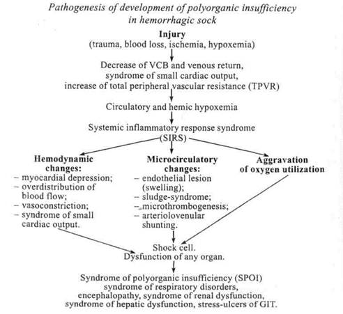
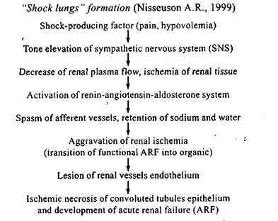
In the development of SPOI three main phases are distinguished: 1. Induction phase that is triggered by different factors (trauma, blood loss, ischemia and infection). These effects convert polymorphonuclear leukocytes and endotheliocytes into the states of "oxygen outburst" the result of which is a chaotic release of cytokins, eicosanoids, mediatory amines and others into a blood flow. 2. A cascade phase is accompanied by development of acute pulmonary lesion, activation of cascades of kallicrein-kinin system, renin-angiotensin-aldosterone system, a system of arachidonic acid, coagulative system and others. 3. A phase of secondary autoaggression is characterized by the utterly On the basis of this it is necessary to single out pathogenesis of "shock kidneys" and "shock lungs" formation.
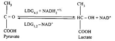
Three stages are singled out in the formation of respiratory distress syndrome (Gattioni L., Bombino M., Pelosi P. et al., 1994). The 1-st stage. Activated leukocytes and thrombocytes are accumulated in capillaries, interstice, releasing, in so doing, prostaglandins, toxic oxygenous radicals and proteolytic enzymes. Lesion of endothelium of pulmonary capillaries and alveolar epithelium leads to plasmorrhagia in the interstice and into alveolar space, atelectasis. The 2-nd stage: interstitial and bronchoalveolar inflammation, proliferation of both epithelium and interstitial cells. The 3-rd stage: interstitial fibrosis that leads to a decrease of ventilation-perfusion ratio, pulmonary hypertension and respiratory hypoxemia. Shock cell: — circulatory and hemic hypoxemia; — ischemia (disturbance of specific and nonspecific functions); — activation of anaerobic glycolysis and inhibition of oxidative decarboxylation of pyruvate;
— decrease of activity of tricarboxylic acid cycle (TCAC) (depression of acetyl-CoA), tissue respiration (inhibition of NAD2+), synthesis of higher fatty acids (HFA), cholesterol (depression of acetyl-CoA) that leads to development of hypoergic state; — lactate acidosis -^ disconnection of oxidative phosphorylation; — inhibition of activity of YJ - Na+ ATP-ase and Ca2+ - Mg2+ - ATP-ase —> cellular hyperhydration (swelling of endotheliocytes) + retention of Ca2+ in cytoplasm (activation of apoptosis, Pg-cascade, disconnection of oxidative phosphorylation, spasm);
— swelling of mitochondria: disconnection of oxydative phosphorylation, depression of tissue respiration, disturbance of shuttle mechanisms functions (glycerophosphate and malat aspartate) -^ increase of redox potential —> Krebtri effect —» decrease of activity of tricarboxylic acid cycle (TCAC); — activation of lipids peroxidation (LPO) and decrease of antioxidant defense; — as a result of swelling of lysosomes a lesion of membrane occurs and release of proteases that leads to autolysis (it is a pathophysiologic basis for decompensatory irreversible shock); — activation of kallicrein-kinin system;
As a finale of shock a cell death occurs: — hypoxic necrobiosis; — free-radical necrobiosis. Дата добавления: 2015-02-05 | Просмотры: 922 | Нарушение авторских прав |


