|
АкушерствоАнатомияАнестезиологияВакцинопрофилактикаВалеологияВетеринарияГигиенаЗаболеванияИммунологияКардиологияНеврологияНефрологияОнкологияОториноларингологияОфтальмологияПаразитологияПедиатрияПервая помощьПсихиатрияПульмонологияРеанимацияРевматологияСтоматологияТерапияТоксикологияТравматологияУрологияФармакологияФармацевтикаФизиотерапияФтизиатрияХирургияЭндокринологияЭпидемиология |
CORRECT ANSWERS
LIST OF REFERENCES A handbook on measures of the first medical aid and prevention of poisonings, injuries and diseases associated with shipment of dangerous cargoes / Ed. by A.A. Lobenko. - Odessa, 1992. - 82 pp. Alternative medicine / Ed. by N.A. Belyakov. - S.Petersburg -Arkhangelsk: North-West Publishing House, 1994. - P. 225-265. Anesthesiology and reanimatology: teaching aid / E. by O.A. Dolina. - M.: Medicine, 1998.-544 pp. Bunyatyan A.A., Ryabov G.A., Manevich A.Z. Anesthesiology and reanimatology. - M.: Medicine, 1984. - P.381-388. Chepky L.P., Zhalko-Titarenko V.F. Anesthesiology and reanimatology. -Kiev: Vysshaya shkola, 1983. - P.292-302. Cohen A.J. Physiologic Concepts in the Management of Renal, Fluid and Electrolyte Disorders in the Intensive Care Unit // Intensive Care Medicine / Edited by J.M. Rippe, R.S. Irwin, M.P Finh, F.B. Cerra. - Vol. I. - Boston -New York - Toronto-London; Little, Brown & Co., 1996. - P.935-950. Cohen A.J., Clive D.M. Acute Renal Failure in the Intensive Care Unit // Ibid.-P. 1000-1023. Extreme and Military Medicine. Collection of test tasks. / V.I. Molchanov, Yu.A. Babushkin, V.V. Kudinov et al. / Reviewer O.N. Spitsin; Crimean State Medical University named after S.I. Georgievsky. - Simferopol: Tavrida, 2002.-263 pp. Intensive therapy of patients with icterohemorrhagic leptospirosis accompanied by polyorganic insufficiency with predominance of acute renal failure / K.Z. Minina, A.A. Titov, LA. Khripachenko et al. // Pain, anesthesia and intensive care. - 1998. - No.3. - P.21-28. Intensive therapy / P.L. Marino, edited by A.I. Martynov. - M.: GEOTAR, Medicine, 1998.-639 pp. Komarov B.D., Shimanko I.I. Positional compression of tissues. - M.: Medicine, 1984.- 176 pp. Kats D., Fridrih P. Volume replacement in the syndrome of polyorganic insufficiency // Current problems of anesthesiology and reanimatology. -Astrakhan-Tramse, 1997. - P.273-275. Medicine of catastrophes: Teaching aid / Ed. by V.I. Molchanov. -Simferopol: Tavrida, 2002. - 308 pp. Nikolayev A.Yu., Milovanov Yu. S. Treatment of renal insufficiency. M.: MIA, 1999.-363 pp. Owen W.F.J. Dialysis Therapy in the Intensive Care Setting // Intensive Care Medicine / Edited by J.M. Rippe, R.S. Irwin, M.P. Fink, F.B. Cerra. -Vol. I. - Boston-New York-Toronto-London: Little, Brown & Co., 1996. -P.1057-1084. Ryabov G.A. Syndromes of critical states. - M.: Medicine, 1994. - 368 pp. Ryabov S.I., Natochin Yu.V. Functional nephrology. - S.Petersburg: Lan', 1997.-304 pp. Shimanko I.I. Lesion of the kidneys in acute exogenic poisonings. M.: Medicine, 1977.-208 pp. Teaching-and-methodical aid on anesthesiology-reanimatology and intensive care of urgent states for students of medical and stomatological faculties, interns, doctors of the faculties of postdiploma education, anesthesiologists-resuscitators. Part II. / Ed. by V.I. Molchanov. - Simferopol, 2000. - 105 pp.
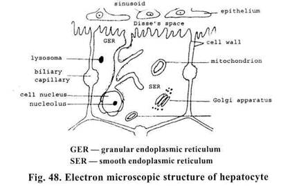
3.6. ACUTE HEPATIC INSUFFICIENCY A liver of the adult man weighs about 1.5 kg (2% of body mass). It consists of the greater right and the less left lobes, as well as of two small lobes — quadrate and caudate lobes. There are 4 species of elements distinguished in the liver: hepatocytes (more than 60% of the cell composition of the liver), reticuloendothelial system (up to 20%), blood and lymphatic vessels, biliary tract. The basic many-sided, versatile work of the liver is performed in hepatocytes that have specific cell organellas for this, discerned in electron microscopy (Fig. 48).
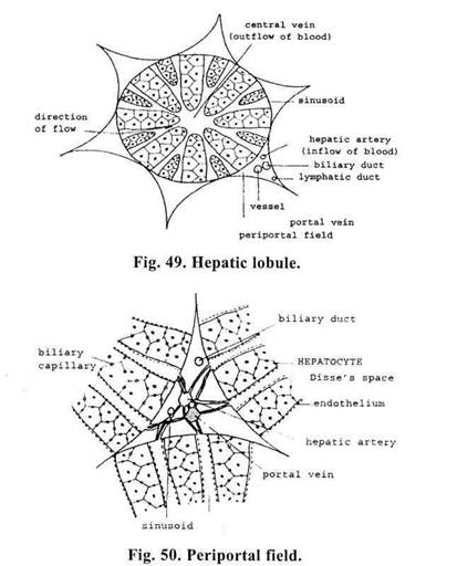
In hepatocyte we distinguish a vascular pole in which endocytosis (capture of substances from the outside and their incorporation into the cells) is performed through microvilli, and a biliary pole where an excretion of substances from the cells (exocytosis) occurs by means of microvilli. The cell membrane for 40-50% of its surface lies adjacent to a sinusoid. A cytoplasmic membrane of this area transfers, actively and selectively, substances from blood into hepatocyte and vice versa. Approximately 30-40% of cell's surface occupies the area through which an exchange of substances is carried out between adjacent hepatocytes. About 10% of cells' surface open into bile passages. There are mitochondria — energy factory of the cell — in the cytoplasm where oxidizing reactions are going on with production of energy from carbohydrates and fatty acids. The important part of the cell is endoplasmic reticulum (cytoplasmic network that confines a cavity of small tubes and vesicles). Through the pores it communicates with the extracellular medium and at the same time with the cell nucleus. A part of reticulum, into the walls of which ribosomes are incorporated, is called granular and the rest — smooth. While proteins are synthetized with the help of ribosomes in the granular endoplasmic reticulum, in its smooth part a detoxication is carried out by way of biotransformation and conjugation of foreign substances (drugs, endotoxins) and metabolytes (bilirubin, urea), thereby their transformation is achieved from liposoluble into water-soluble substances,as well as synthesis of fatty acids. It is also possible that endoplasmic reticulum is responsible for a transport of different substances into the cell, from the cell and inside the cell. It is connected with laminar Golgi apparatus concentrating and packing substances to be transported. So, carbohydrate-and-protein complexes are obtained in the form of glycoproteids and glucosaminoglycanes. Lysosomes are membraneous vesicles rich in hydrolases that carry out splitting of large molecules of cellular and extracellular origin. Such fate waits for bacteria penetrating a hepatocyte. Lysosomes are closely connected with laminar Golgi complex, participate in bile secretion. Sharp pH changes of the medium and lesion of lysosomes may lead to activation of hydrolases, acid phosphatase in particular, and to cells' destruction. Enzymes come out into a vascular bed where their activity sharply increases. Cell nucleus contains genetic material in the form of DNA. There are nucleoli in the nucleus that contains RNA transfering the information to a cytoplasm where, in accordance with the obtained information, new substances are synthetized, for example, enzymes. A cell nucleus is surrounded with a double membrane. The external nuclear membrane is a component part of the endoplasmic reticulum through which a nucleus is connected with the cell environment. A nuclear membrane has numerous pores that ensure a migration of substances between nucleus and protoplasm. Every cell is surrounded with three-layered cellular membrane, so-called basal membrane that separates a cell medium from an extracellular one. An anatomical unit of the liver is hepatic hexagonal lobule (Fig. 49) formed by the bars of hepatocytes that are arranged radially around a central vein (origin of hepatic veins) and separated by slits called sinusoids. Between hepatic lobules are wedge-shaped periportal fields (Fig. 50) in which small branches of portal vein, hepatic artery, lymphatic vessels and small biliary ducts pass through. A hepatic blood flow makes up 100 ml/min/100 g., that is 25% of the cardiac output. Hepatic artery supplies 25% of hepatic blood flow and 45-50% of oxygen, and portal vein -r» 75% of blood flow and 50-55% of oxygen, in spite of the fact that the portal vein has already given back a portion of oxygen to the digestive organs and spleen. The pressure in hepatic artery is equal to systemic arterial pressure, and in the portal vein — 7-10 mm Hg. From minute
branches of v. porta and arterioles of a. hepatica with the pressure of about 40 mm Hg blood enters sinusoids. Presinusoid (precapillary) and postsinusoid (postcapillary) sphincters regulate alternating blood flow from v. porta and a. hepatica preventing excessive variations of pressure in sinusoids. Having /?-adrenoreceptors the system of pre- and postsinusoid sphincters take part in defence response of the organism to a decrease of cardiac output, e. g., in shock. Thus, in the course of hemorrhage the liver can add up to 500 ml of blood to the system of blood circulation. Affection of the liver reduces its sensitivity to catecholamines and thereby a reaction of centralization of circulation is disturbed. In prolonged bleeding necrobiotic changes of hepatocytes may develop. Particularly sensitive to oxygen deprivation are the centers of lobules where, even in the norm, the cells are poorly supplied with oxygen compared to the periphery of lobule. In sinusoids optimal conditions are created for the exchange of gases, nutrients and waste products between the cells and mixed blood. Sinusoids function as capillaries in other areas of the body. From them blood is supplied to central veins (Fig. 49). Sinusoids are covered with very loosened layer of the endothelium, through numerous fenestrae (windows) that are in their diameter less than formed elements of blood, plasma, from sinusoid, easily penetrates the cavity between endothelium and hepatic cells (Disse's space) and wash hepatocytes. On the side directed to Disse's space hepatocyte has numerous very narrow passages that ensure the exchange between the cell and Disse's space. In case of need they can dilate increasing the intensity of exchange. Kupffer star mast cells are arranged in sinusoids, the former protect hepatocytes from foreign particles coming into blood, phagocyting and digesting some products of blood coagulation, contrast substances, artificial colloids that are applied for volumotherapy, etc. The sides of hepatocyte, adjacent to each other, form biliary capillaries: in the middle into which bile is secreted (Fig. 48). Thus, the walls of biliary capillaries are nothing less than the walls of hepatic cells. They turn into biliary ducts that, in their turn, are communicated with large intrahepatic biliary duots. The right and left lobes have major ducts that form together a common duct — ductus hepaticus that after its confluence with ductus cysticus bears the name — choledochus. It enters duodenum on papilla duodeni. Дата добавления: 2015-02-05 | Просмотры: 970 | Нарушение авторских прав |